


- 23
- 54


- 12
- 67
close-up version of this post
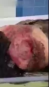
https://watchpeopledie.tv/h/medical/post/11928/archive-mmc-after-an-unknown-flesheating


- 9
- 21
As promised, here are the pictures of my recent cranial MRI. But first, conext:
I have not yet had the corresponding doctor's appointment to discuss findings so I am not going to be commenting on the images apart from a brief explanation in regards to my already diagnosed illnesses. I picked two images that I find are representative of the results, though I do have 92 total images.

This is an image in sagittal view (t2), you can see no dawson's fingers, a telltale sign of MS which I was expecting to see here and throughout. My MS diagnosis has been established through MRI of the spinal canal, where I do have lesions although I didn't think to ask for the pictures at the time. I am not going to speculate on what this means exactly, this is just further context.

This is an image in axial view(t2), again no lesions expected in MS are visible. Note that my brain does have a slanted ass though. (For my fellow med fans, does that count as midline shift?)
Also a ping because you've been bugging me @Owenreal


- 3
- 33
The attached image demonstrates cobblestoning of intestine. It is seen in Crohn's disease. The condition can also be seen in:
Hirschsprung disease
candida esophagitis
eosinophilic gastritis
Brunner gland hyperplasia
duodenitis
nodular lymphoid hyperplasia
Reference:
https://radiopaedia.org/articles/cobblestone-appearance-hollow-viscera
Image via:
https://radiopaedia.org/articles/cobblestone-appearance-hollow-viscera


- 3
- 37
The papillary muscles are specialised muscles located in the ventricles of the heart. They attach to the cusps of the atrioventricular valves (also known as the mitral and tricuspid valves) via the chordae tendineae or heart strings (The white cords in the given photo) and contract to prevent inversion or prolapse of these valves during systole or ventricular contraction.


- 3
- 24
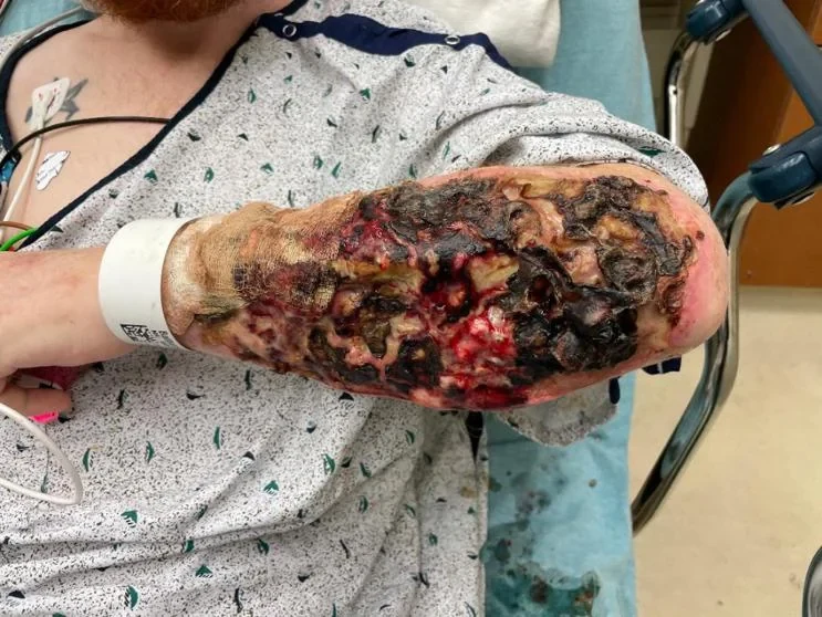
A 35-year-old male presented to the emergency department with bilateral forearm wounds. His past medical history was significant for untreated hepatitis C. He was unemployed and homeless. He reported injecting xylazine-laced fentanyl into his bilateral forearms. A physical exam revealed bilateral full-thickness skin wounds from the wrist to the proximal forearm over the ulnar border measuring [Figure 1]. The wounds appeared cavernous with undulated regions of eschar. Further, examination revealed exposed muscle and tendon bilaterally and some exposed muscle of the dorsal forearm compartments. His hand function was preserved.
The patient was admitted to the hospitalist and initiated on broad antibiotic treatment with intravenous vancomycin and piperacillin/tazobactam. His wounds were washed and covered at the bedside, and he underwent surgical debridement in the operating room the following day. Intraoperatively, the eschar layers were removed sharply with a knife until a bleeding surface was encountered. Exposed muscle layers were removed with a curette. The ulna had an intact periosteum. Large-volume irrigation was used to clean the wounds. A negative pressure wound therapy device was fitted to the bilateral forearms. The sponge was changed twice over six days. On a postoperative day seven, the patient was returned to the operating room. Granulation tissue was observed over the ulna, and the wounds appeared clean. The surgeon covered the area with a split-thickness skin graft taken from the thigh. A negative pressure wound dressing was placed over the skin graft. The patient left the hospital against medical advice (AMA) two days following the second surgery and did not follow up in the surgeon's office.
Approximately nine months later, the patient returned to the emergency room with a similar, smaller wound on the left forearm. On exam, both previous skin grafts had healed well, and the new wound was adjacent to the volar border of the skin graft and measured 8x4cm. The wound exhibited a cavernous appearance but did not have exposed muscle. The next day, surgical debridement was performed, followed by negative pressure wound therapy. The day after surgery, the patient left AMA with no follow-up reported since.


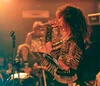
 Posting medical gore is my thing
- 1
- 13
Laparoscopic Cholecystectomy is a minimally invasive surgical procedure to remove the gallbladder. The procedure is performed through small incisions in the abdomen, using special instruments and a camera to visualize the internal organs. Here is a step-by-step explanation of the procedure:
Anesthesia: The patient is given general anesthesia and is asleep during the procedure.
Making Incisions: The surgeon makes several small incisions in the abdomen, usually less than an inch long.
Inserting the Laparoscope: A laparoscope, a thin tube with a camera and light at the end, is inserted through one of the incisions to allow the surgeon to see the inside of the abdomen.
Creating a Pneumoperitoneum: Carbon dioxide gas is pumped into the abdomen to create a space for the surgeon to work and get a better view of the organs.
Disconnecting the Gallbladder: The gallbladder is carefully detached from the liver and bile ducts, and any stones present are removed.
Removing the Gallbladder: The gallbladder is removed through one of the incisions, usually through the umbilicus (belly button).
Closing the Incisions: The incisions are closed with small stitches or surgical glue and covered with a dressing.
Recovery: After the procedure, the patient is monitored for any complications and is usually able to go home the same day or the next day. Pain and discomfort can be managed with pain medication. Most patients can return to normal activities within a few days.
Laparoscopic Cholecystectomy is a safe and effective way to remove the gallbladder and can provide relief from symptoms such as abdominal pain and indigestion. It is important to follow the surgeon's post-operative instructions and to attend follow-up appointments as advised to ensure a smooth recovery.

- 1
- 19
An 18-year-old man with hemophilia A presented to the otolaryngology clinic with a 4-day history of throat discomfort, painful swallowing, and difficulty moving his tongue.
He had moderately severe hemophilia A with a baseline factor VIII activity level of 2 to 5% (reference range, 70 to 150).
He administered recombinant factor VIII as needed for bleeding episodes and had started doing so 2 days before this presentation for a right neck hematoma. He recalled no recent neck or throat trauma, coughing, or choking.
On physical examination, a resolving right anterior neck ecchymosis was seen (Panel A).
Laryngoscopy showed multiple hematomas on the right oropharyngeal wall (Panel B, asterisk), lingual epiglottic surface (Panel B, arrowheads),
and aryepiglottic fold (Panel C, arrows).
The factor VIII activity level was 9%. In patients with hemophilia, pharyngeal or laryngeal bleeding may manifest as sore throat, odynophagia, vocal changes, or tongue discomfort. Factor replacement therapy should be administered urgently and empirically if there are diagnostic delays to avoid the progression of bleeding to airway compromise. The patient continued to receive recombinant factor VIII, and at follow-up 10 days later, his symptoms and hematomas had resolved.


- 9
- 29


 In Hospital


- 10
- 54
I'm so confused as of what's going on.
Might not be a CW but I'll be safe
Please feel free to move this if it's in the wrong flair or reposted 


Top Poster of the Day:
 reichsfuhrer
Deaths Today: 0
Current Registered Users: 2,107,378
 BROWSE EFFORTPOSTS
SITE GUIDE
PING GROUPS
BROWSE EFFORTPOSTS
SITE GUIDE
PING GROUPS
Medical

Aspiring medical student? Or maybe just have an interest in human anatomy? Here you will find everything from autopsies to surgeries to photos from medical journals. If it happened in the operating room or the morgue, it probably belongs here.
 Slavshit
 Sandshit